Diversity of Klebsiella Surface Antigens O and K
*Note: This hosting platform doesn't permit italicization of the genus name in this article header.
01 — Objective: To strengthen a grant proposal by creating tailored, comprehensive visual representations of the structural diversity of Klebsiella surface antigens, while indicating their localization within the bacterial cell membrane.
02 — Material:
Many Klebsiella species can be found in low numbers in a healthy human microbiome; still, opportunistic agents within the genus, including members of the Klebsiella pneumoniae species complex, Klebsiella oxytoca, and several others, represent long-standing threats to immunocompromised and otherwise vulnerable individuals. These conditional pathogens are causative agents of a range of healthcare-associated infections, including nosocomial pneumonia, catheter-associated urinary tract infections (CAUTIs), surgical site and wound abscesses, and fatal sepsis.
In recent years, emerging hyper-virulent (hvKp) and multi-drug
resistant (MDR/cKp) Klebsiella strains have also begun to
affect immunocompetent individuals, and now pose a mounting worldwide
health concern. These infectious Klebsiella species possess an
arsenal of virulence factors that contribute to pathogenicity and
assist in evasion of the host immune response.
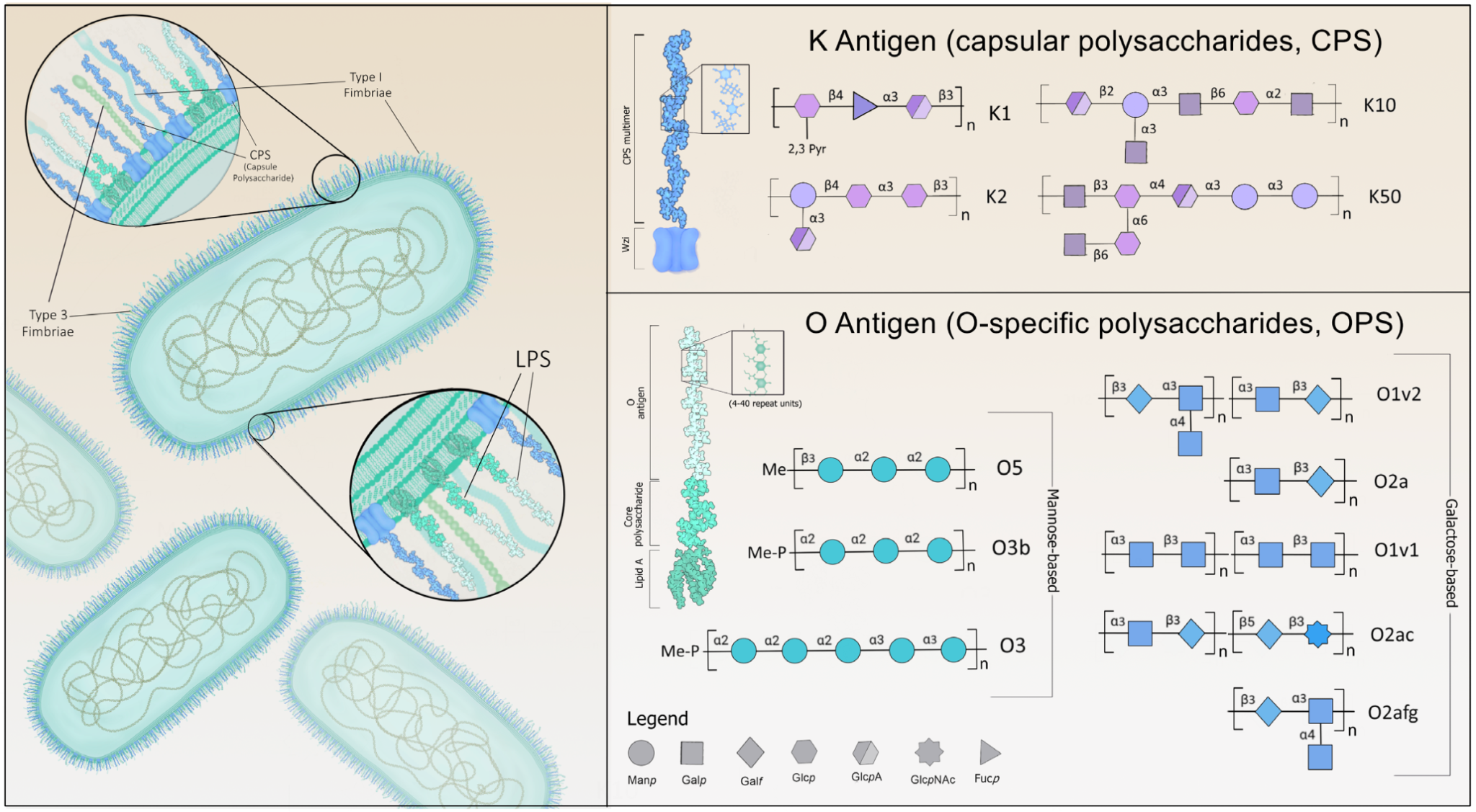
Klebsiella K
antigens (capsular polysaccharides) are a diverse group of
surface molecules containing 77-79 serologically-characterized
structural variations. Broadly, K antigens help protect the bacteria
from the host immune response at multiple levels, helping the
bacteria to avoid detection, evade cell-mediated immunity, and
block innate immune signaling cascades. The K antigen group
has a hand in reducing engulfment of the bacteria by epithelial
cells, avoiding phagocytosis by macrophages and neutrophils,
interrupting NF-kB signaling, and triggering cell death and
cytotoxicity in host cells. In hvKp strains specifically, key K
antigens, such as K1 and K2 (Figures 1 and 2), help the opportunistic
pathogen evade clearance by the host by conferring increased
resistance against two of these facets of the immune response:
complement-mediated killing and phagocytosis. Less
well-understood,
serologically-identified
variations, including K5, K20, K54, K57, and K64, are also associated
with a hypervirulent phenotype.
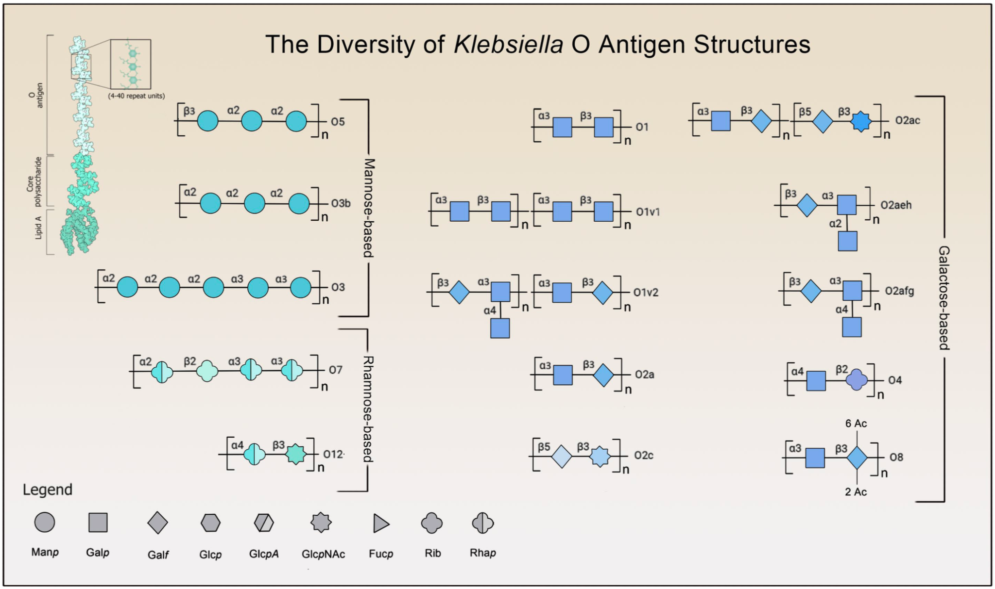
The O-specific polysaccharides (OPS), or O antigens, are a group containing around 9-13 structural variations, although O1, O2, and O3 (Figure 2) are identified in about 80% of infections. In tandem with CPS, O-specific polysaccharides help counter the host immune response by suppressing TLR-signaling and facilitating resistance to complement-mediated killing through interference with C3b deposition. The O1 variant is often seen in hvKp infections along with K1 and K2; at present, O1, O2, O3, and O5 are represented across emerging MDR/cKp strains. Altogether, the structure of these factors varies meaningfully across distinct strains, contributing in part to the rapidly developing pathologies seen in MDR, hvKp, and convergent (MDR-hvKp) species.
Over the past two decades, multi-drug resistance has been increasingly observed as a regular and concerning feature of severe human infection by opportunistic and hyper-virulent Klebsiella species. The spread of drug resistance forebodes an eventual narrowing of treatment options for Klebsiella infection, while, in parallel, an increase in hyper-virulence abets a concurrent widening of the susceptible population. The role of surface antigenic variation in individuating MDR and HV-specific pathology warrants further characterization of these rapidly-evolving virulence factors. Exploration of novel therapeutic agents and vaccines targeting these antigens denotes a promising strategy to preempt this rising global health threat.
The 3D, space-filling drawings of the O and K antigens were illustrated from representative structures available in the database K-PAM: Klebsiella species serotype predictor and surface antigens modeler, which contains further references within. The symbolic representations of the diversity of structures were obtained both from this database and from Clarke et al (2018). Opoku-Temeng et al. (2019) and Wang et al. (2024) provided insight into the localization of both antigen classes, and nomenclature was derived from Kaptive 2.0.
03 — Impact: Directly supported successful acquisition of funding that initiated a new research trajectory for the client’s laboratory. The figures served as lasting reference material for ongoing project documentation and presentations.
Post a comment